One of the most common questions I am asked is about skin lumps and whether or not they are dangerous.
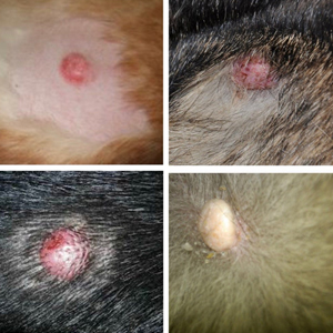
As you will see in the photograph the lumps look fairly similar, but 2 are totally benign, 1 is potentially malignant and 1 is malignant. Can you tell which is which? I can’t just by looking!
At college we’re taught the importance of not using guesswork to diagnose a skin lump, but instead to test them. David Grant MBE, BVetMed, CertSAD, FRCVS, a renowned vet dermatologist, who spends all day looking at lumps once only got 50% right by simply looking at them – if he couldn’t tell by looking at a lump what it was definitively then neither can the rest of us!
So how do we tell what they are? How do we know if they need removing or if they can be left. Or if they need close monitoring.
Initially the visual appearance is important eg: is there redness, ooze, infection, irritation or ulceration. Is it growing slowly or quickly. Does it grow outwards (like a nipple) or within the skin. Can an edge be felt, is it soft, hard, fluid, lumpy?
Then we have to go past what we can see with the naked eye and down to the cellular level. To do this we need a sample from the lump.
The simplest test is an FNA (fine needle aspirate) because it is relatively painless and results can be obtained quickly. However, it isn’t always accurate or diagnostic. The vet aims the needle into the mass to withdraw some cells but cannot see where the tip of the needle is.
Sometimes the cells withdrawn are from just the right area and a diagnosis is gained. Other times the cells may be dying in part of the lump or they won’t suck up into the needle. Even with these limitations it is one of the most useful and frequently performed tests in my practice.
Secondly we can get a biopsy from the lump. Sometimes this means removing the whole lump and sending it all for analysis. On other occasions we may take a piece of tissue out the lump and stitch it up. This is useful for lumps in areas that have very little skin to close the wound. This type of biopsy can help us with planning what to do next.
If the results come back as a slow growing, non-invasive mass that is unlikely to spread then we may leave it. Or we may still remove it (especially if it annoys the pet) but we know that we don’t have to cut away a lot of normal tissue around it.
However, if the results show a malignancy we may decide to do other testing before removal. This is called staging and usually entails blood testing, radiographs, ultrasonography or other advanced imaging like CT scans.
It is also helpful to know how much of a margin we need around the mass. Basically this means cutting the tumour out, but trying to make sure that no microscopic cells are left behind, by taking away some normal tissue too. This is all sent off to the laboratory to find out if we succeeded in getting clean borders.
Although we do see a lot of skin tumours that need removal many of them carry a good prognosis, especially if we diagnose them early. So don’t panic if you see a lump but do get it checked out.